
If your healthcare professional suspects that you may have a brain tumor, a series of advanced tests and medical procedures are required to confirm the diagnosis. Early and accurate diagnosis plays a critical role in determining the best treatment plan and improving survival outcomes.
This comprehensive guide explains:
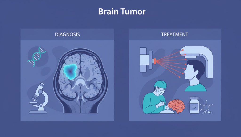
- Brain tumor diagnostic tests
- MRI, CT scan, PET scan & biopsy
- Brain tumor grading system
- Survival rates and life expectancy
- Treatment options including surgery, radiation, chemotherapy & targeted therapy
- Prognosis factors
How Is a Brain Tumor Diagnosed?
When symptoms such as persistent headaches, seizures, vision changes, memory loss, or personality changes appear, doctors begin a structured evaluation process.
1. Neurological Examination
A neurological exam is often the first step in diagnosing a brain tumor.
This exam evaluates how well different areas of your brain are functioning. It may include testing:
- Vision and eye movements
- Hearing ability
- Balance and coordination
- Muscle strength
- Reflexes
- Sensation
- Memory and concentration
Although a neurological exam cannot directly detect a brain tumor, it helps doctors determine which part of the brain may be affected. For example:
- Speech difficulty may indicate involvement of the left hemisphere.
- Balance problems may suggest cerebellar involvement.
- Weakness on one side may point to motor cortex issues.
This exam guides doctors toward the most appropriate imaging test.
2. CT Scan (Computed Tomography)
A Head CT scan uses X-rays to create detailed images of the brain.
Why CT Scans Are Important:
- Widely available
- Quick results
- Useful in emergency situations
- Detects bleeding, swelling, and large tumors
CT scans are often the first imaging test performed when someone has severe headaches, sudden neurological symptoms, or suspected brain injury.
If the CT scan shows abnormalities, your doctor will usually recommend a more detailed brain MRI.
3. Brain MRI (Magnetic Resonance Imaging)
MRI is the gold standard for detecting brain tumors.
MRI uses powerful magnets and radio waves to produce highly detailed images of brain tissue.
Why MRI Is Superior:
- Better soft tissue contrast
- Detects smaller tumors
- Clearly shows tumor boundaries
- Differentiates tumor from healthy brain tissue
Often, a contrast dye (gadolinium) is injected into a vein before the MRI. This contrast helps:
- Highlight tumor cells
- Distinguish tumor from swelling
- Identify aggressive tumor areas
Advanced MRI Techniques
Modern neuro-oncology uses specialized MRI techniques:
Functional MRI (fMRI)
- Maps brain areas responsible for speech, movement, and thinking
- Helps surgeons avoid damaging critical areas
- Essential for surgical planning
Magnetic Resonance Spectroscopy (MRS)
- Measures chemical composition inside tumor cells
- Identifies tumor type
- Helps distinguish tumor recurrence from radiation damage
Magnetic Resonance Perfusion
- Measures blood flow within tumor tissue
- Identifies highly active tumor regions
- Assists in treatment planning
4. PET Scan (Positron Emission Tomography)
A brain PET scan detects metabolic activity within tumor cells.
During the procedure:
- A radioactive tracer is injected into the bloodstream.
- Tumor cells absorb more tracer than normal cells.
- Highly active tumors appear brighter on images.
PET Scan Is Most Useful For:
- Fast-growing tumors (like glioblastoma)
- Evaluating tumor recurrence
- Detecting aggressive tumor behavior
Slow-growing benign tumors may not appear clearly on PET scans. Not every patient requires a PET scan; your doctor will decide based on your case.
Brain Biopsy: Confirming the Diagnosis
Imaging tests can strongly suggest a brain tumor, but only a biopsy can confirm the diagnosis.
What Is a Brain Biopsy?
A biopsy involves removing a small sample of tumor tissue for laboratory analysis.
There are two main types:
1. Surgical Biopsy
Performed during tumor removal surgery.
2. Stereotactic Needle Biopsy
Used when surgery is risky or the tumor is deep within the brain.
In stereotactic biopsy:
- A small hole is drilled into the skull.
- A thin needle is guided into the tumor using MRI or CT imaging.
- Tissue samples are collected.
The procedure is performed under anesthesia, so you won’t feel pain.
Risks of Brain Biopsy:
- Bleeding
- Infection
- Brain swelling
- Temporary neurological symptoms
Despite risks, biopsy is essential for accurate tumor classification.
Laboratory Testing of Tumor Tissue
After biopsy, the tissue sample is analyzed in a laboratory.
Pathologists examine:
- Whether cells are cancerous (malignant) or noncancerous (benign)
- How abnormal the cells appear
- How fast they are dividing
- Genetic mutations and DNA changes
These findings determine the tumor grade and guide treatment decisions.
Brain Tumor Grading System
Unlike other cancers, brain tumors are not staged (Stage 1–4). Instead, they are classified by grade.
Grade 1 Brain Tumor
- Slow-growing
- Cells look similar to healthy cells
- Often benign
- Better prognosis
Grade 2 Brain Tumor
- Slightly abnormal cells
- Slow but can recur
Grade 3 Brain Tumor
- Clearly abnormal cells
- Faster growth
- Malignant
Grade 4 Brain Tumor
- Highly aggressive
- Rapid growth
- Cells look very different from normal
- Worst prognosis
The most aggressive example is glioblastoma (Grade 4).
Brain Tumor Prognosis
Prognosis refers to the likely outcome of the disease.
Factors affecting prognosis include:
- Tumor type
- Tumor grade
- Tumor location
- Genetic mutations
- Patient age
- Overall health
- Whether tumor can be completely removed
Brain Tumor Survival Rates
Overall Survival Statistics:
- 1-year survival rate: ~40%
- 5-year survival rate: ~19%
- 10-year survival rate: ~13%
These numbers combine all tumor types and grades.
Life Expectancy After Brain Metastasis
Brain metastasis occurs when cancer spreads from another part of the body to the brain.
Average survival after brain metastasis:
- Typically 3–12 months
- With stereotactic radiosurgery (SRS): 12–15 months
- 3-year survival: About 5%
Outcome depends on:
- Original cancer type
- Number of brain tumors
- Treatment response
- Overall health
Life Expectancy for Grade 4 Brain Cancer
Glioblastoma:
- Age 65+: 6–9 months
- Age 18–44: 20 months
- Age under 17: 15 months
Diffuse Midline Glioma (DMG):
- Less than 1 year
Anaplastic Astrocytoma (Grade 3):
- Around 25 months
Anaplastic Oligodendroglioma:
- Up to 15 years
- 20% survive 5 years or less
Common Causes of Death from Brain Tumors
Brain tumors can cause death due to:
- Increased intracranial pressure
- Brain swelling
- Bleeding (hemorrhage)
- Infections
- Compression of vital brain centers
Brain Tumor Treatment Options
Treatment depends on:
- Tumor type
- Tumor grade
- Location
- Size
- Patient health
- Personal preferences
1. Surgery for Brain Tumor
The goal of surgery is complete tumor removal when possible.
Craniotomy
Most common procedure:
- Section of skull removed
- Tumor removed
- Skull replaced
Awake Brain Surgery
Patient awakened during surgery:
- Ensures speech and motor areas aren’t damaged
Endoscopic Brain Surgery
- Minimally invasive
- Often used for pituitary tumors
- Accessed through the nose
Risks of Surgery:
- Infection
- Bleeding
- Vision or hearing loss
- Brain tissue damage
Partial tumor removal (subtotal resection) may still relieve symptoms.
2. Radiation Therapy
Radiation uses high-energy beams to destroy tumor cells.
External Beam Radiation
- Given 5 days per week
- 2–6 weeks duration
Whole Brain Radiation
Used for multiple metastases.
Proton Therapy
- More precise
- Protects healthy tissue
- Useful for children
Side Effects:
- Fatigue
- Hair loss
- Memory issues
- Scalp irritation
3. Stereotactic Radiosurgery (SRS)
Despite its name, no surgical incision is made.
Types:
- Gamma Knife
- LINAC (CyberKnife)
- Proton radiosurgery
Highly focused radiation kills tumor cells in one or few sessions.
4. Chemotherapy
Uses strong drugs to kill cancer cells.
Given:
- Orally
- IV injection
- Directly into brain tissue during surgery
Side effects:
- Nausea
- Hair loss
- Fatigue
- Weakened immunity
5. Targeted Therapy
Targets specific genetic mutations in tumor cells.
Advantages:
- More precise
- Fewer side effects
- Personalized treatment
Not all tumors qualify for targeted therapy.
Recovery After Brain Tumor Treatment
Rehabilitation may include:
Physical Therapy
- Improve movement and strength
Occupational Therapy
- Regain daily function
Speech Therapy
- Improve speech and communication
Cognitive Therapy
- Improve memory and thinking
Children may require educational support.
Final Thoughts
A brain tumor diagnosis can feel overwhelming, but modern medical advances have significantly improved diagnostic accuracy and treatment outcomes.
Early detection through neurological exams, CT scans, MRI, PET scans, and biopsy ensures proper classification and grading.
Treatment options such as surgery, radiation therapy, chemotherapy, radiosurgery, and targeted therapy offer hope and extended survival for many patients.
If you experience persistent neurological symptoms, seek medical evaluation promptly. Early diagnosis can improve both survival and quality of life.